Business & Tech
3-D Imaging Technology Is Revolutionizing Dental Surgery
Technology provides more accurate information prior to dental implant procedure.
While 3-D may be the latest, greatest thing in the realm of television and movie entertainment, a local dental surgeon is finding a more practical use for the technology.
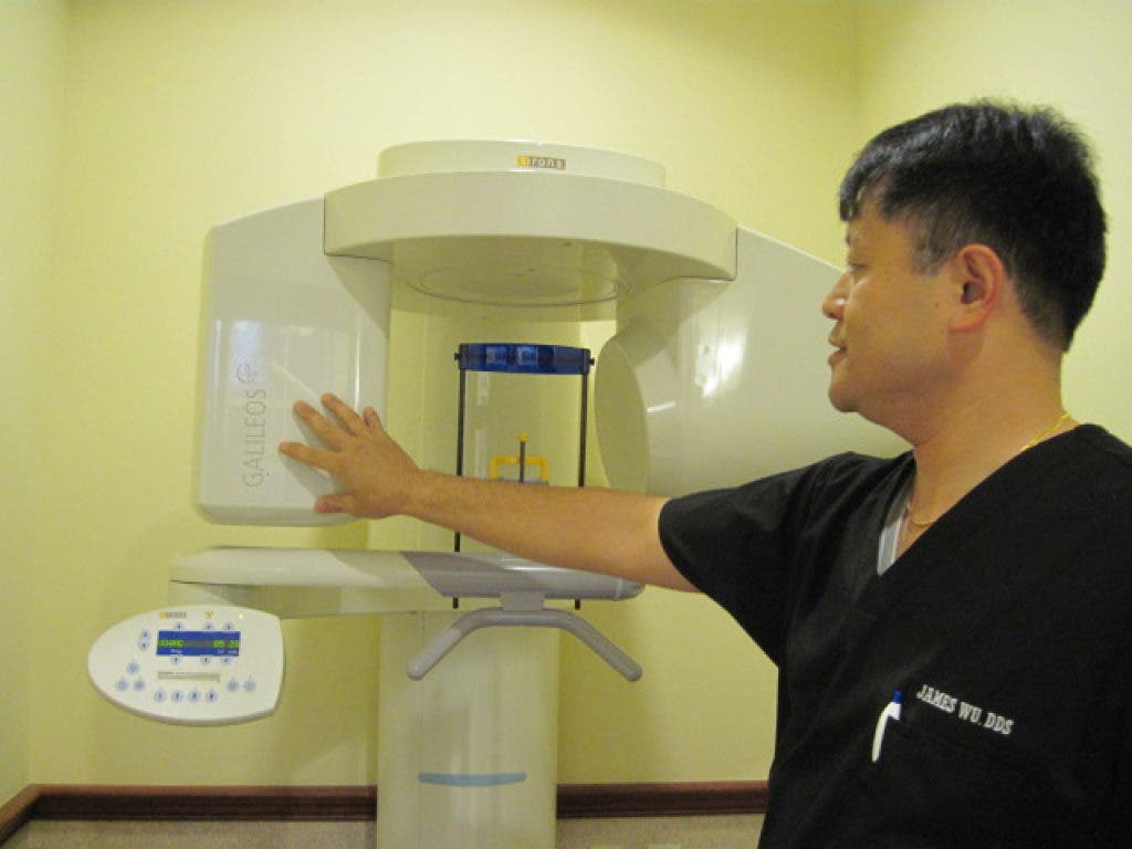
When practice to 1201 Main St. in 2009, he also invested in the 3-D Imaging Cone Beam Scanner for the purposes of dental implant treatments.
“Dental implants are basically tooth roots that can be placed into the jaw area when a patient has a missing tooth,” said Wu, who shares his practice with Dr. Daniel Jeong. “When the new implant root has healed to the jawbone, then teeth can be connected on top.
Interested in local real estate?Subscribe to Patch's new newsletter to be the first to know about open houses, new listings and more.
"In order to place a dental implant and have it be successful long-term, you need to have good bone structure around the implant root just like a natural tooth.”
Wu also said that the size of the tooth missing determines the size of the implant needed.
Interested in local real estate?Subscribe to Patch's new newsletter to be the first to know about open houses, new listings and more.
“We need to know that the patient has enough bone in the area…in terms of width and height for the patient to be able to have a dental implant placed," he said.
Wu said that by taking a simple look into a patient’s mouth, dentists can get a good idea of bone structure, and two dimensional X-Rays, the common media for most dentists today, can determine the width and height, but not depth. These two-dimensional X-Rays can be deceiving. Wu said that as a result, surgeons have gone in to do a dental implant treatment only to find that the bone isn’t deep enough.
“The problem with that is that now, you’ve committed to a surgery that you cannot perform, so now you’re altering your treatment," Wu explained. "What the 3-D imaging allows us to do is to know all that information up front … what the width of the bone is, what the height, what the depth is so we can be certain when we go in there to perform treatment that it can be performed … that there are no surprises going in.”
Wu says that 3-D imaging started picking up in the early 1990’s, but back then, the 3-D imaging was done using standard, medical grade CT scans. They gave off much higher levels of radiation than is used in scanning today. The type of scanning that Wu uses is called “Cone Beam CT scanning.”
“The Cone Beam Scanner is one point of radiation that fans out in a cope shape, and takes the image with just the cone beam, whereas the medical CT scanner does images in layers … then you put the layers together," said Wu. "It’s not one continuous scan. That’s why there’s much more radiation when you take the medical grade CT scan. The reason that the (Cone Beam Scanner) has much less radiation is because it’s all done in one scan.”
Wu states that the levels of radiation given off by the Cone Beam Scanner are as much as 60 to 70 percent less radiation than the medical grade CT scans.